Anatomy, structure and size of the kidney
In this article we will describe the size, anatomy and structure of the kidney. It uses quite alot of ‘doctors language’, sorry [“yeah right!” CKDEx Ed].
Size
In adults, the normal kidney is 10-14 cm long in males and 9-13 cm long in females, 3-5 cm wide, 3 cm in antero-posterior thickness and weighs 150-250 g. The left kidney is usually slightly larger than the right.
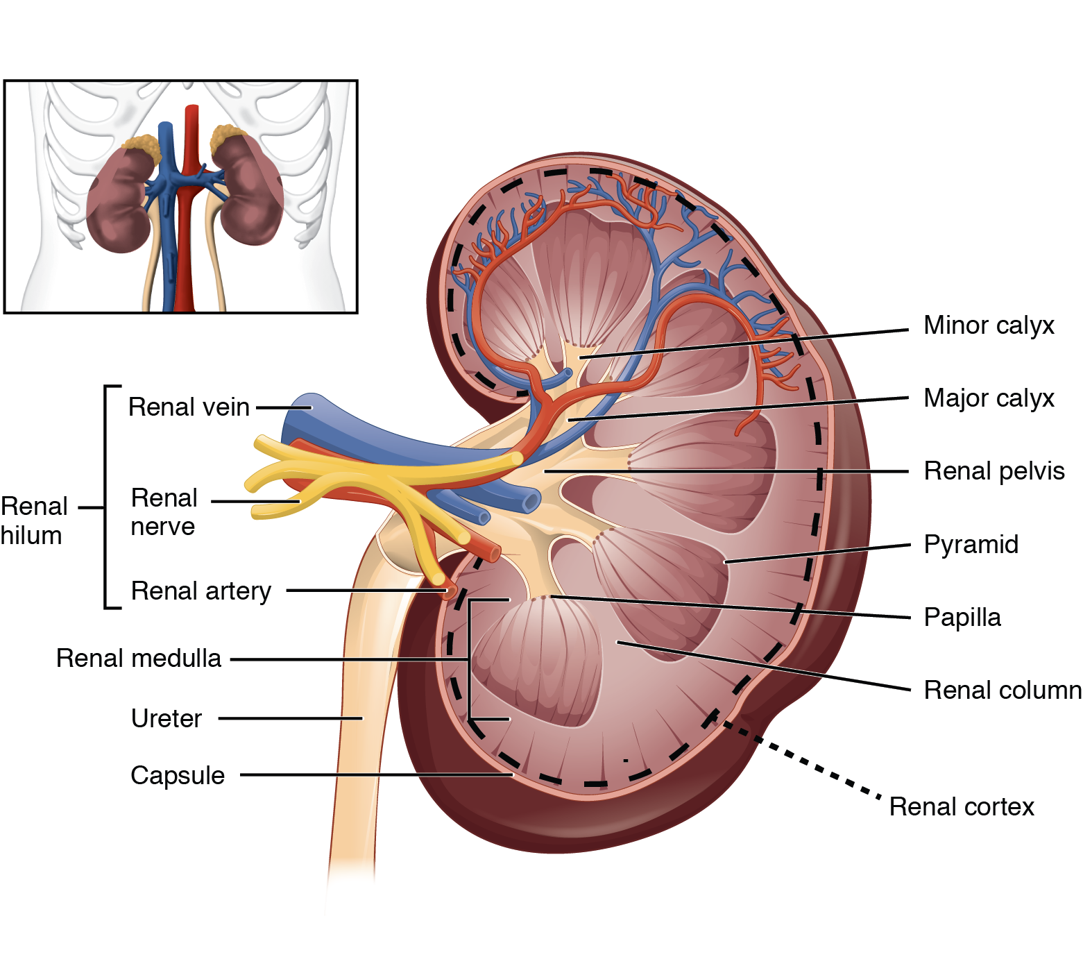
Diagram courtesy of Oregon State University.
Anatomy and structure
The kidney is bean-shaped with a superior and an inferior pole, anterior and posterior surfaces, and lateral and medial borders. This is fancy-docs language for the kidney being a ‘3D thing’! The midportion of the kidney is called the midpole – another fancy word [“thanks for that!” Ed].
The kidney has an outer fibrous capsule, which is surrounded by perirenal fat.
Internally, the kidneys have an intricate and unique structure. The kidney itself can be divided into:
- Renal parenchyma (outer area), consisting of an outer renal cortex and inner medulla
- Renal sinus (inner area) containing the hilum, renal pelvis, calyces, renal blood vessels, nerves, lymphatics and perirenal fat.
The cortex extends into the medulla, dividing it into triangular shapes – these are known as renal pyramids (10-14 of those) [“nothing to do with the Pyramids of Egypt”. Ed]. The apex of a renal pyramid is called a renal papilla. Each renal papilla is composed of structures called minor calyces (singular = ‘calyx’; means a cup-like cavity), which collect urine from the pyramids.
Several minor calyces merge to form a major calyx (2-3 of these). Urine passes through the major calyces which unite to form the renal pelvis, a flattened and funnel-shaped structure. From the renal pelvis, urine drains into the ureter, which transports it to the bladder for storage.
The medial (inner) side of each kidney is marked by a deep fissure, known as the renal hilum. This acts as a gateway to the kidney. Normally the renal vessels and ureter enter/exit the kidney via this structure – with the pelvis/ureter posterior (behind) the blood vessels, and the renal vein anterior (in front of) the renal artery.
Language notes
Note 1. A ‘hilum’ is a ‘small thing’ in Latin; and in biology it has come to mean a depression or fissure where structures such as blood vessels and nerves enter an organ
Note 2. ‘Pelvis’ means a ‘basin’ in Latin, from the Greek word pelex (‘helmet’) and pelike (‘goblet’ or ‘bowl’).
Summary
We have described the size, anatomy and structure of the kidney. We hope it has been helpful.
Other resources
Where are the kidneys located?
Are the kidneys the same size?
Review article: anatomy of kidneys (Soriano, 2023)
https://www.youtube.com/watch?v=Qz8UZ1ROSqU&t=1s
Last Reviewed on 22 May 2024