
How does the glomerulus work?
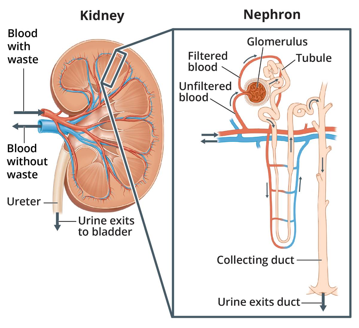
A glomerulus is a tiny filter in the kidney. There are a million in each kidney. In other words the kidney is not one big filter, it is a million tiny ones.
The first step in making urine is to separate the liquid part of your blood (plasma), which contains all the dissolved minerals and waste products, from your blood cells. This is the function of the glomerulus which is a microscopic filter (sieve), that is constantly filtering your blood.

Stretched out from end to end, the nephrons (that includes the glomeruli and tubules) are about 5 miles (8 kilometers) long.
What pushes blood through the glomerulus?
Blood that is about to be filtered enters the glomerulus, which is a scrunched up ball of blood capillaries (the smallest blood vessels) – like a ball of string or worms. The glomerulus is nestled inside a cup-like sac located called a glomerular (or Bowman’s) capsule. Glomerular capillaries have small pores in their walls, just like a very fine mesh sieve.
The glomerulus is sandwiched between two arterioles – afferent arterioles deliver blood to the glomerulus, whilst efferent arterioles carry it away. Constriction of efferent arterioles as blood exits the glomerulus provides resistance to blood flow, preventing a pressure drop; which could not be achieved if blood were to flow into venules, which do not really constrict.*
The two arterioles change in size to increase or decrease blood pressure in the glomerulus. In addition, efferent arterioles are smaller in diameter than afferent arterioles. As a result, pressurised blood enters the glomerulus through a relatively wide tube, but is forced to exit through a narrower tube.
Together, these unique features plus the fact that your heart is supplying your kidneys with over a litre of blood per minute (around 20% of its output) maintain a high glomerular capillary pressure; and the filtration function of the kidney occurs, regardless of fluctuations in blood flow.
For example, the sympathetic nervous system can stimulate the efferent arteriole to constrict during exercise when blood flow to the kidney is reduced.
Glomerular capillary wall

.jpg)
Diagram and electron microscope image of glomerular capillary wall consisting of three layers: endothelium, basement membrane, and epithelium
The physical characteristics of the glomerular capillary wall determine what is filtered, and how much is filtered into the glomerular capsule. Working from the inside out, the capillary walls are made up of three layers:
-
Endothelium – this has relatively large pores (70-100 nanometers in diameter), which allows solutes, proteins and fluid to pass through, but not blood cells
-
Glomerular basement membrane (GBM) – this membrane is fused to the endothelial and epithelial layers. Its job is to prevent proteins from being filtered out of the bloodstream
-
Epithelium – this layer consists of specialised cells called podocytes. These cells are attached to the basement membrane by ‘foot processes’ (pedicels; see diagram above). They wrap around the capillaries, but leave slits between them, known as filtration slits. A thin diaphragm between the slits acts as a final filtration barrier before the fluid (now called ultrafiltrate) enters the glomerular space.
What happens next?
After the ultrafiltrate exits the glomerulus (where it has accumulated in Bowman’s space), it flows into a thin tube called the proximal convoluted tubule (PCT). This is the first part of a long tube called the renal tubule.
Together each glomerulus and linked tubule are called a nephron, as shown in the diagram at the top of the page.
As it moves, substances that are needed and some water are reabsorbed (pulled back) through the tube wall into adjacent capillaries. This reabsorption of vital nutrients from the filtrate is the second step in urine creation.
The tubules eventually join up and form the ureter which takes the fluid (now called urine) to the bladder. From there it is removed from the body when you have a wee.
Summary
We have described how a glomerulus works. Yes, its an amazing little filter isn’t it? We hope it has been helpful.
Last Reviewed on 22 May 2024