What is a nuclear medicine renal (kidney) scan?
In a nuclear medicine renal scan (or ‘renogram’), pictures are taken of radioactive substances going into the kidneys from the bloodstream, inside the kidneys, and into the bladder through the ureters (that join the kidneys to the bladder).
A nuclear medicine renal scan uses radiopharmaceuticals (radioactive substances) injected into a vein, usually in the arm, to provide clear images of the kidneys taken with a special camera called a gamma camera. They are done in part of an x-ray department.
There are three main types.
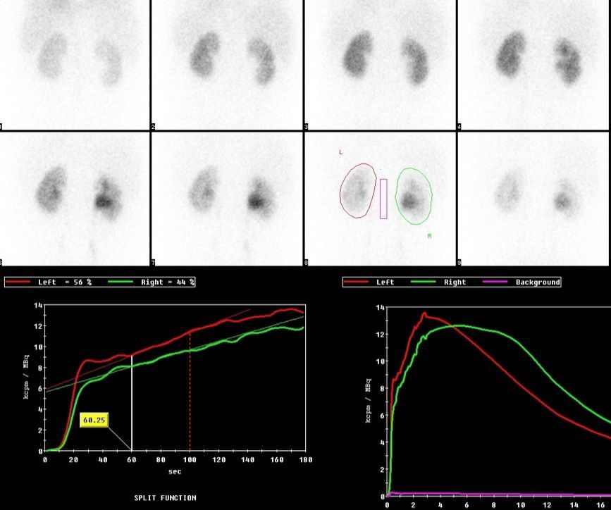
DTPA and MAG3 scans – are similar and look at the functions of the kidney. They are used to look for obstruction to the kidney urine drainage system, and blood supply to the kidney. They can also tell you the ‘split function’, i.e. if there are two kidneys, what is the contribution of each to making effective urine.
The scan can be carried out using one of two different radiopharmaceuticals – Technetium-99m DTPA (diethylene triamine pentaacetic acid) or Technetium-99m MAG3 (mercaptoacetyletriglycine). They are similar substances, but MAG3 gives significantly clearer images in some patients, particularly very young children and those patients with poor kidney function.
DMSA scan – is used to look at the structure of the kidney. For example they can diagnose renal scarring from chronic infection, or renal infarction (dead kidney tissue from a blockage to the blood supply to an area of the kidney). It uses a radioactive substance called Technetium-99m DMSA.
What do these scans look like?
Yes, they are both vague and/or complicated. This is why specialist x-rays doctors are required to interpret them.
Isotope GFR test
An isotope GFR test is used to assess the function of your kidneys very accurately, through a series of blood tests. It uses a radioactive substance called chromium-51 EDTA.
This procedure gives a more accurate measurement of the glomerular filtration rate (GFR; i.e. total kidney function) than an estimated GFR (eGFR), which is based on the blood creatinine level – the worse the kidney function, the higher is the creatinine, and lower the eGFR.
It is useful when the doctor is not sure if you have CKD or not. This is especially true in muscular people (larger muscles increase the creatinine level, not just CKD); when the eGFR is borderline low, which could be a sign of mild CKD.
What are the risks of a renal scan?
Nuclear medicine scans are safe. The radioisotope exposes you to less radiation than an x-ray. The small amount of radiation exposure is primarily in the kidney area. It passes from your body naturally within 24 hours.
The low doses of radiation used in nuclear medicine procedures don’t have a connection to any long-term negative effects.
Even though the radiation exposure is minimal and short-term, tell your doctor if you’re pregnant or think you might be pregnant. Also, tell your doctor if you’re breastfeeding to ensure that there’s no contamination of your breast milk.
Unlike intravenous dyes, radioisotopes carry a very low risk of an allergic reaction. Allergic reactions to radioisotopes are possible but rare. A renal scan is a good option if you’ve had a reaction to the contrast dye used in X-rays of the urinary system.
Last Reviewed on 2 September 2023