An ultrasound scan uses ultrasound, or high-frequency sound waves, to create an image (picture) of part of the inside of your body, such as your kidney.
It is used in many other branches of medicine to look at other internal organs (like the heart when it is called an echocardiogram, liver, or pelvic organs), or a baby inside a pregnant woman.
You cannot hear ultrasound waves, but it can be picked up by a special ultrasound machine. Ultrasound waves pass through fluid (liquid) and soft tissues, and bounce back (like an echo) from more solid structures.
Why will I need an ultrasound?
The renal (kidney) ultrasound is one of the three key tests need, when doctors are investigating someone with CKD. It is done for two reasons:
- To check you have two kidneys, sometimes as a prelude to a renal (kidney) biopsy
- To determine the cause of CKD, e.g. polycystic kidney disease (PCKD).
The other two key tests are: (1) blood creatinine (and eGFR that comes from it) level, and (2) protein levels in the urine (‘urinary albumin-to-creatinine ratio, or uACR’).
What are the risks and complications?
An ultrasound scan is very safe, because sound waves do not harm the body. The scan does not hurt.
What happens during an ultrasound?
The ultrasound scan normally takes place in the x-ray department of your hospital. A doctor or a sonographer (a specialist technician trained in the use of ultrasound), performs the test. Your doctor uses the information to help make a diagnosis.
The scan usually takes about 30 minutes. This is the process:
- You lie down on your back. A gel is put on your skin – this may feel a bit cold
- Then the operator may get you to roll on your side or front:
- A small handheld device called a ‘transducer’ is moved over your skin – the gel helps it move smoothly. This transducer is attached to a computer and a monitor
- Pulses of ultrasound are sent through the transducer and into the body – you cannot feel these
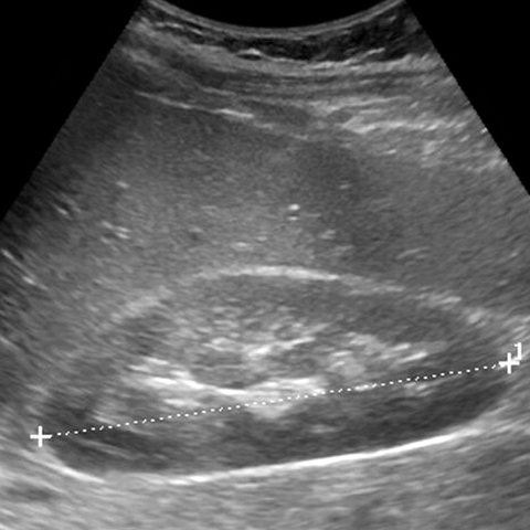
- These sound waves bounce back from the structures inside the body (like an echo). They are displayed as an image on the monitor (screen), that looks like this:

A normal kidney looks on an ultrasound. The technician is measuring the length of it
- The operator will also have a look at your liver, and other internal organs, to check these are OK
- Sometimes, an ultrasound scan can show moving images, like a video.
Last Reviewed on 16 September 2023
